
6 Exercises for Individuals With Scoliosis Exercise cannot straighten a scoliotic spine, but the right movements can meaningfully reduce pain, improve muscle balance, and support better posture. These six exercises are appropriate for many individuals living with scoliosis, though any new exercise program should be discussed with your spine specialist before you begin. Living with…
Read more
News
Most often diagnosed during childhood or adolescence, scoliosis can vary widely in severity and long-term impact. One of the most common questions patients and families ask is whether scoliosis is genetic. To answer that and other frequent questions, this article explains what is known about the inheritance of scoliosis, how it develops, and what it…
Read more
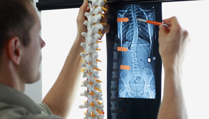
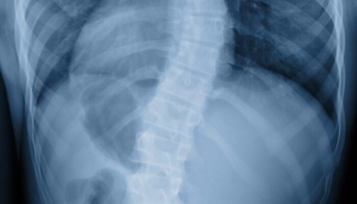
Scoliosis is a condition of the spine in which the backbone curves to the side in a way that is different from the normal alignment. Instead of running straight down the middle of the back, the spine forms a curve that looks like the letter C or the letter S when viewed from behind. While…
Read more

Recovering from a herniated disc can feel slow and uncertain. For many patients, the gradual return of comfort, function, and mobility can be hard to recognize day by day. Understanding the typical signs that your spine is healing can help you feel more confident about recovery and know when it may be safe to resume…
Read more

A herniated disc occurs when the soft inner material of a spinal disc pushes through the outer layer, sometimes irritating nearby nerves. This can cause back or neck pain, numbness, tingling, or weakness in the arms or legs depending on the location of the disc. Fortunately, there are a range of treatment options, starting with…
Read more

While a herniated disc can be painful and disruptive, many cases resolve without surgery. Knowing the signs, understanding your options, and seeking care early can help you get back to normal activity faster and reduce the chance of recurrence. Back pain is one of the most common medical complaints in the United States. For many,…
Read more

Brain hematomas are more complex—and more dangerous—than many people realize. From the risk factors posed by common medications like blood thinners to life-threatening pressure that can physically shift the brain, these conditions demand swift medical attention. Some develop slowly, others strike without warning, and all require a keen awareness of symptoms and follow-up care. Georgia…
Read more

Though often used interchangeably, brain hematomas and brain hemorrhages are not the same. A hemorrhage refers to active bleeding, while a hematoma is a collection of blood that typically forms after the bleeding has begun. This article explains their relationship and outlines common types of brain hemorrhages—including epidural, subdural, subarachnoid, intracerebral, and intraventricular—highlighting how each…
Read more

Brain hematomas occur when blood vessels in or around the brain rupture, leading to dangerous pressure buildup. Symptoms include severe headaches, nausea, confusion, and vision changes. Diagnosis typically involves CT or MRI scans, and treatment depends on severity. Acute cases often require emergency craniotomy surgery, while chronic hematomas may be treated with less invasive burr…
Read more

A subdural hematoma is a life-threatening condition caused by bleeding in the membranes surrounding the brain, typically due to head trauma. Unlike superficial bruises, subdural hematomas do not show on the skin but instead cause severe headaches, nausea, slurred speech, vision changes, dizziness, weakness, memory loss, or personality changes. Symptoms can appear immediately, within hours,…
Read more